A 73 year old man presented with headache and progressive weakness affecting the left arm which slowly progressed over 3 hours.
![]()
![]()
![]()
![]()
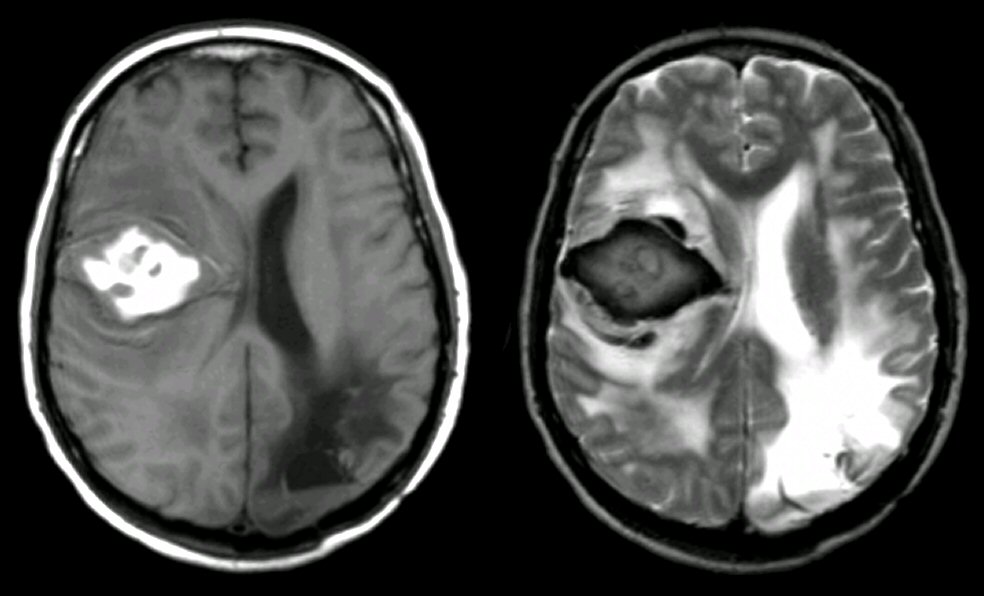
Axial MRI scans: (Left) T1-weighted; (Right) T2-weighted. Note on T1, there is an area of hyperintense signal in the right frontal lobe. The same area on T2 is dark with a surrounding bright signal. This is the characteristic picture of a subacute early (3-7 days) hemorrhage on MRI. The hyperintense signal on T1 with the hypointense signal on T2 is is the pattern seen with intracellular methemoglobin. The bright surrounding signal on T2 is vasogenic edema. In this case, there is also old encephalomalcia in the left occipital lobe from a previous stroke. The findings of blood on MRI are complex and depend on timing. To learn more, review the powerpoint slide show, Blood on MRI: Time-dependent Changes. In this case, the hemorrhage was due to hypertension.
Revised05/02/06.
The Electronic Curriculum is copyrighted 1998, Case Western
Reserve University
School of Medicine.